
Whenever you need a highly detailed picture of your insides, then the best way to get it through imaging tests. Formally known as imaging studies, these are tests that do exactly what we’ve mentioned and are very accurate.
Their use mostly comes when diagnosing various forms of cancer and other potentially life-threatening diseases. These studies can be done in multiple ways using multiple techniques for different purposes; mostly related to uncovering different forms of cancer.
There are many different techniques out there, and the most commonly used are radioactive particles, magnetic fields, X-rays, sound waves, etc.
Each technique is very effective in providing the doctor or medical expert with a highly detailed imagery of the insides of a patient. These images give back crucial information of stuff such as organs, tissue, cells, and lots of others.
As we mentioned earlier, certain techniques achieve this in different ways, but all are very accurate and precise.
What Do Imaging Studies Reveal
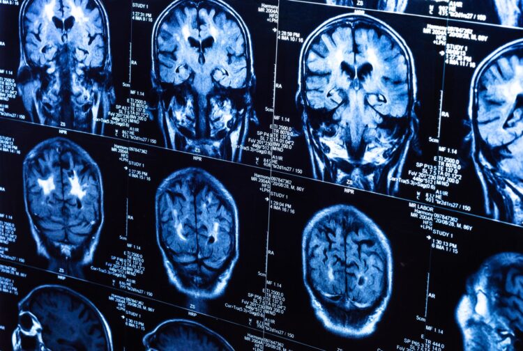
Since medicine is improving and perfecting with each passing year, the use of imaging studies proves instrumental in the fight against cancer.
Their use comes mostly in the detection and prevention area, where patients can use any number of these studies to get an accurate representation of their insides.
Imaging studies are an improved technique for finding cancer cells than endoscopic tests. Endoscopic tests rely on the doctor seeing the cells through a lens that is sent through a tube into patients’ insides.
This lens is effectively a camera that provides feedback through a monitor. But we no longer need to use of such studies or testing, since imaging studies are a better, more complete alternative to uncovering cancer cells.
Also, imaging studies are the best way to uncover cancer before it takes a huge toll on a patient’s body and health. Endoscopic tests will only show cancer cells once they appear and start to do damage to a patient. Imaging studies, on the other hand, can detect cancer cells around any part of the body, even if they’re well hidden within tissue or organs.
This is called screening and is highly recommended that everyone should do it on a regular basis.
Through screening, we also make it possible to pinpoint the exact location of a particular cancer or tumor and even see it as it is growing. We can also see the extent of the tumor or cancer and use the right means to treat it.
So, with all that said, what are the types of imaging study techniques?
Well, let’s find out.
Computed Tomography
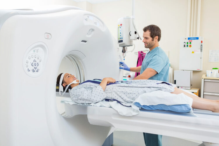
This technique is used to screen and scan only a single organ or a part of the body. It uses a light beam that sends the information to a computer where it generates an image of the organ or part of the body being scanned.
This image is very hard to obtain so doctors inject a dye to the patient so the image could be clearer and easier to obtain and generate.
The finalized image comes in the form of an x-ray image.
This type of screening, along with the others we’ll mention, can be provided to you at any facility that specializes in imaging studies. For more information, make sure to visit wispecialists.com.
MRI
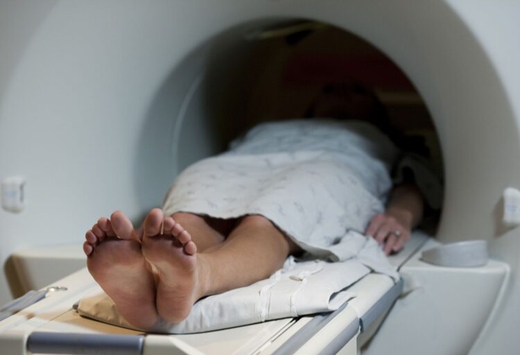
Magnetic resonance imaging does the same exact thing as computed tomography but uses magnetic fields to obtain the images.
MRI is especially precise and accurate when trying to uncover cancerous or tumor cells to a specific part of the body. This includes the liver and the nervous system (the brain). This technique is especially common in today’s medicine and is all possible through the discovery of the magnetic field in science.
With that said, MRI uses a magnetic field onto a laying patient while they’re inside a huge device called a scanner. The machine does use some radiation to help obtain the images. MRI uses the radiation with the magnetic field to align the hydrogen atoms in our tissues. Once everything is done, the atoms are aligned back into their original state and the machine gives us a 3D image of our body. Again, same as computed tomography, we can inject dyes for obtaining a clearer image.
Mammography
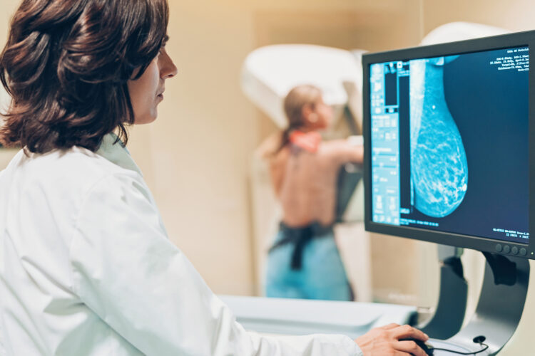
Mammography is solely used to examine the breasts and uncover cancerous or tumor cells into this particular part of the body. Mammography does this through the use of x-rays and is very effective in detecting all kinds of anomalies into the breasts.
Mammography is mostly used for women in uncovering cancer or tumor to the breasts. But unlike other radiation-based techniques, this one uses lower radiation levels due to safety concerns. With all that said, mammography is perfectly safe and excellent when needing a well-rounded image of a woman’s breasts.
Nuclear Scan
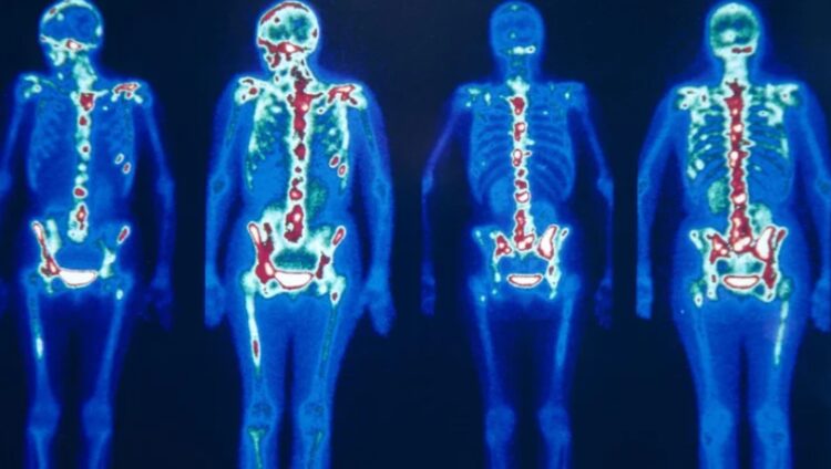
This is a very different study technique than others we’ve mentioned because it is far less commonly used. Nuclear scanning or scintigraphy also uses low levels of radiation (much like mammography) to uncover cancer or tumor cells to a patient’s body.
The scan is done by giving the patient tracers that are absorbed through the tissue. These tracers go through the body and flag any parts that have cancerous or tumor cells.
Nuclear scans are a popular tool to uncover various other issues. Their use is primarily in the prevention and uncovering of cancer, but others include thyroid scans, liver scans, bone scans, and spleen scans.
PET
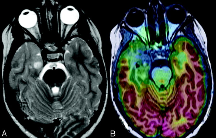
PET or positron emission tomography is yet another type of tomography technique that uses radiation for the base of it.
This particular type uses an atom that emits a particle called a positron. This atom is actually radioactive by nature and the positron can be absorbed by multiple means.
Most PET scans are used to include the likes of brain imaging studies with a focus on uncovering cancer. Other areas also include the lungs and liver, breasts, colon, ovaries, and others.
These were some imaging studies techniques that we can use to uncover cancer and tumor to a patient’s body.


